A sudden fall, a hard tackle, or even an awkward reach can leave you clutching your shoulder in intense pain. Few injuries feel as alarming as a shoulder slipping out of place, and the moment it happens, most people know something is seriously wrong.
Shoulder dislocation happens when the head of the upper arm bone moves out of its socket. This injury usually affects the main shoulder joint and is considered a medical emergency because nearby nerves and blood vessels can be harmed. Fast medical care lowers the risk of complications, supports proper healing, and helps restore safe movement. With the right treatment plan, many people return to their normal activities without lasting problems.
In this guide, you will learn what shoulder dislocation is, why it happens, how it is treated, and what you can do to prevent it.
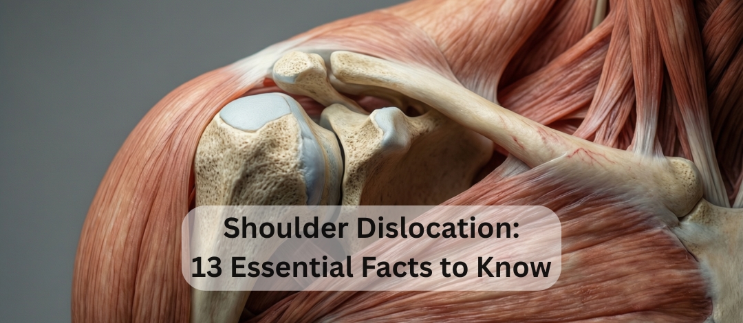
Shoulder Dislocation: 13 Essential Facts to Know

A shoulder dislocation is the displacement of the humeral head from the glenoid fossa, meaning the ball of the upper arm bone no longer sits properly in its socket.
The injury most commonly affects the glenohumeral joint, the primary joint responsible for the shoulder’s wide range of motion. Because this joint is highly mobile, it is also more vulnerable to instability.
Doctors treat shoulder dislocation as a medical emergency due to the risk of neurovascular damage. Prompt care helps prevent long-term weakness, numbness, or circulation problems.
Types of Shoulder Dislocation
Shoulder dislocations are classified by direction and severity. Understanding the difference helps guide treatment and recovery expectations.
Based on Direction
These describe where the humeral head moves after leaving the socket.
- Anterior dislocation:
- Makes up about 90–95% of cases
- The humeral head shifts forward and slightly downward
- Often caused by the arm being forced backward while rotated outward
- Posterior dislocation:
- Occurs in roughly 2-4% of cases
- Commonly linked to seizures or electrical injuries
- Can be harder to detect during early exams
- Inferior dislocation (Luxatio Erecta):
- Rare but dramatic
- The arm becomes locked overhead
- Requires urgent medical attention
Based on Severity
Severity reflects how far the joint surfaces separate.
- Complete dislocation: Total loss of contact between the bones
- Subluxation: Partial displacement with some joint contact remaining
Anatomy Involved
The shoulder relies on a delicate balance of bones, soft tissues, and stabilizing structures.
Bones involved:
- Humerus
- Scapula, which forms the glenoid socket
- Clavicle
Key stabilizers:
- Glenoid labrum that deepens the socket
- Joint capsule that surrounds the joint
- Glenohumeral ligaments for structural support
- Rotator cuff muscles that control movement
Neurovascular structures at risk:
- Axillary nerve, the most commonly affected
- Axillary artery, which supplies blood to the region
Damage to these structures is why fast evaluation matters.
Causes and Mechanism of Injury

Most shoulder dislocations result from strong external forces, though some develop from repeated strain.
Common causes include:
- Traumatic injuries such as falls, sports collisions, or vehicle accidents
- Forced abduction combined with external rotation, a classic anterior pattern
- Direct force to the back of the shoulder leading to posterior displacement
- Repetitive instability in athletes who perform overhead motions
- Congenital ligament laxity that allows excessive joint movement
Even a single high-impact event can stretch or tear stabilizing tissues.
Risk Factors
Some people face a higher chance of shoulder dislocation due to lifestyle or physical traits.
- Previous dislocation, which weakens joint stability
- Participation in contact sports
- Younger age groups with higher recurrence rates
- Naturally hypermobile joints
- Weak shoulder stabilizing muscles
Addressing these factors early can lower the risk of repeat injuries.
Clinical Presentation
Recognizing the warning signs helps ensure faster treatment.
Symptoms
People often report intense discomfort and loss of control.
- Severe shoulder pain
- Inability to move the arm
- A sensation that the shoulder has “popped out”
Signs
Visible clues usually confirm that something is wrong.
- Obvious deformity
- Flattened appearance of the shoulder muscle
- Arm held in a fixed or guarded position
- Muscle spasms
- Possible numbness along the outer shoulder, suggesting axillary nerve involvement
Any suspected dislocation should be evaluated immediately.
Diagnosis
Accurate diagnosis confirms the injury and reveals associated damage.
Physical Examination
Clinicians begin with a careful hands-on assessment.
- Inspection for abnormal shape
- Palpation to locate the humeral head
- Neurovascular checks to ensure proper nerve function and blood flow
Imaging
Imaging provides clear confirmation.
- X-ray: Verifies the dislocation and checks for fractures
- CT scan: Useful for complex injuries
- MRI: Detects soft tissue damage such as labral or rotator cuff tears
Immediate Management
Early care focuses on safety, pain control, and restoring alignment.
- Immobilize the arm to prevent further injury
- Provide pain relief and muscle relaxation
- Perform a closed reduction, done only by trained medical professionals
- Repeat neurovascular checks after reduction
- Confirm proper alignment with an X-ray
Attempting to reposition the shoulder without training can worsen the injury.
Treatment
Treatment plans vary depending on severity, patient age, and recurrence risk.
Non-Surgical Care
Many first-time dislocations respond well to conservative treatment.
- Sling immobilization for about 2–6 weeks
- Ice therapy and anti-inflammatory medication
- Gradual physiotherapy to rebuild strength
Surgical Options
Surgery may be recommended when instability persists.
Common indications:
- Recurrent dislocations
- Labral tears such as a Bankart lesion
- Significant bone defects
- Young athletes with ongoing instability
Procedures include:
- Arthroscopic stabilization
- Open repair when additional support is needed
Your orthopedic specialist will guide the best approach.
Rehabilitation
Rehabilitation restores strength, flexibility, and confidence in the joint.
- Phase 1: Immobilization and pain control
Allows tissues to begin healing while reducing inflammation. - Phase 2: Passive range-of-motion exercises
A therapist gently moves the arm to prevent stiffness. - Phase 3: Active strengthening
Focuses on the rotator cuff and scapular stabilizers. - Phase 4: Functional and sport-specific training
Prepares the shoulder for daily tasks and athletic demands.
Skipping rehab greatly increases reinjury risk.
Complications
While many people recover well, complications can occur.
- Recurrent instability
- Axillary nerve injury
- Rotator cuff tears
- Bankart lesions
- Hill-Sachs lesions
- Persistent shoulder stiffness
- Post-traumatic arthritis
Early treatment helps reduce these outcomes.
Prognosis
The outlook for shoulder dislocation is generally positive with proper care.
Most patients regain strong function, especially when treatment begins quickly. However, recurrence is more common in people under 25 due to higher activity levels and tissue elasticity.
Early rehabilitation plays a major role in long-term success, improving strength and helping restore full movement.
Prevention
Although not every injury is avoidable, smart habits protect the shoulder.
- Strengthen rotator cuff and scapular muscles
- Use proper sports technique
- Wear protective gear when appropriate
- Address instability before it worsens
Consistency in training is one of the best defenses against repeat dislocations.
Conclusion
Shoulder dislocation is painful and disruptive, but with prompt care, structured treatment, and dedicated rehabilitation, most people recover well. Understanding the causes, recognizing the symptoms, and taking preventive steps can make a meaningful difference in both healing time and future joint health.